In the course "Diverse Brain" we aimed to innovate on how people communicate and interact with science. We believe that through the use of 3-dimensional exhibition pieces we can not only better capture the attention of the casual onlooker but also are able to communicate more direct and intuitive. Our focus has been the brain, as it is not only incredibly fascinating to us but has also been a major focus of pop-sci for quite some time. Our challenge was to highlight various topics about the brain and clear up common misconceptions. A common one would be the notion of a "left brain" for logical thinking and "right brain" for creative thinking. In this course we co-educated neuroscience, computer science as well as students from many other disciplines from Berlins major universities to go beyond the limits of specialization and benefit from as many perspectives as we could.
Neuroplasticity
Cora-Lorraine Sachs Psychology (FU Berlin)
Nazlican Göksu Tekdogan Computational Sciences (FU Berlin)
Jakob Schmock Computer Science (FU Berlin)
Jonas Heinemann Computer Science (FU Berlin)
Michael Migacev Computer Science (FU Berlin)
While many people believe the brain cannot recover after brain damage, neuroplasticty proves otherwise. Dendritic remodeling, axonal sprouting, and neurogenesis play crucial role in the recovery process.
In this piece we playfully highlight some aspects of neuroplasticity and simulate a healing brain by placing the correct tiles into the correct cutouts. We primarily focus on 3 parts of neuroplasticity:
1. Dendritic remodeling involves changes in the dendrites which are branches of neurons that receive information from other neurons. This process helps neurons adapt to new functions and rebuild damaged circuits. [1]
2. Axonal sprouting occurs when neighboring neurons extend their axons to establish new connections with damaged brain regions. That helps in restoring lost neural paths and functional connections. [2]
3. New neurons can be generated in certain brain regions. Newly generated neurons collaborate with existing connections, and contribute to restoring cognitive functions such as memory and learning. Whether neurogenesis happens on a scale relevant to healing and upholding neural function is still debated. [3]
References:
[1]: RP Stroemer, TA Kent, and CE Hulsebosch. Synchronous neuronal activity is a signal for axonal sprouting after cortical lesions in the adult. The Journal of Neuroscience, 22(14):6062–6070, July 2002.
[2]:: Gregory Yiu and Zhigang He. Axonal regeneration: Underlying molecular mechanisms and potential therapeutic targets. Biomedicines, 10(12):3186, 2022.
[3]:: Dong Sun, Raymond J Colello, William P Daugherty, Taejung H Kwon, Michael J McGinn, and C Edward Dixon. Neurogenesis in adult human brain after traumatic brain injury. Journal of Neurotrauma, 28(4):763–774, 2011.
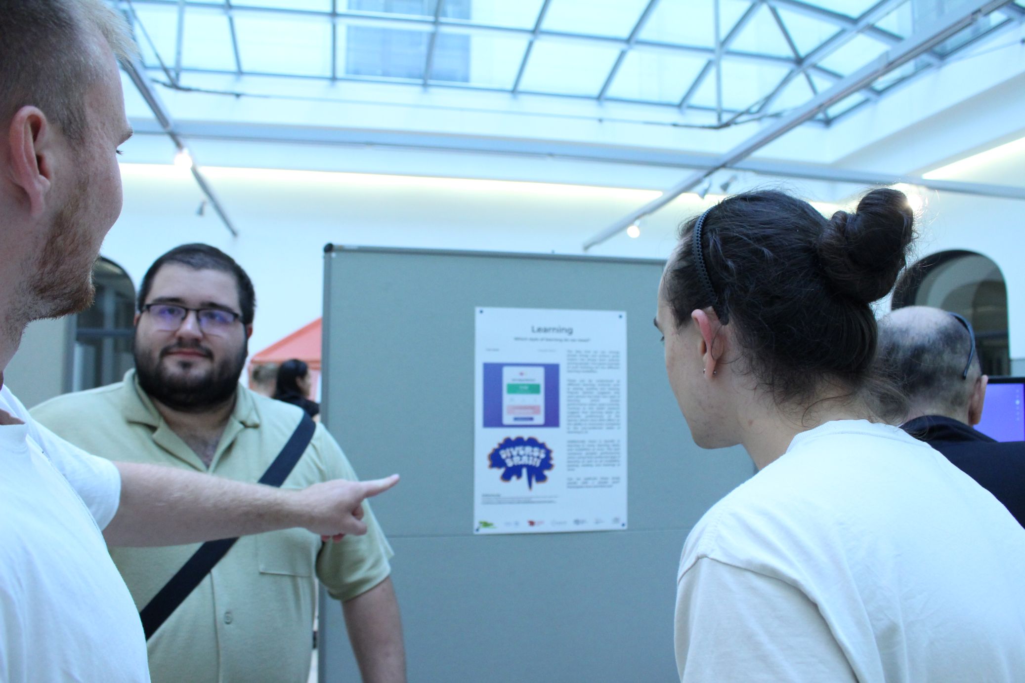
Learning
Paul Nelde Computer Science (FU Berlin)
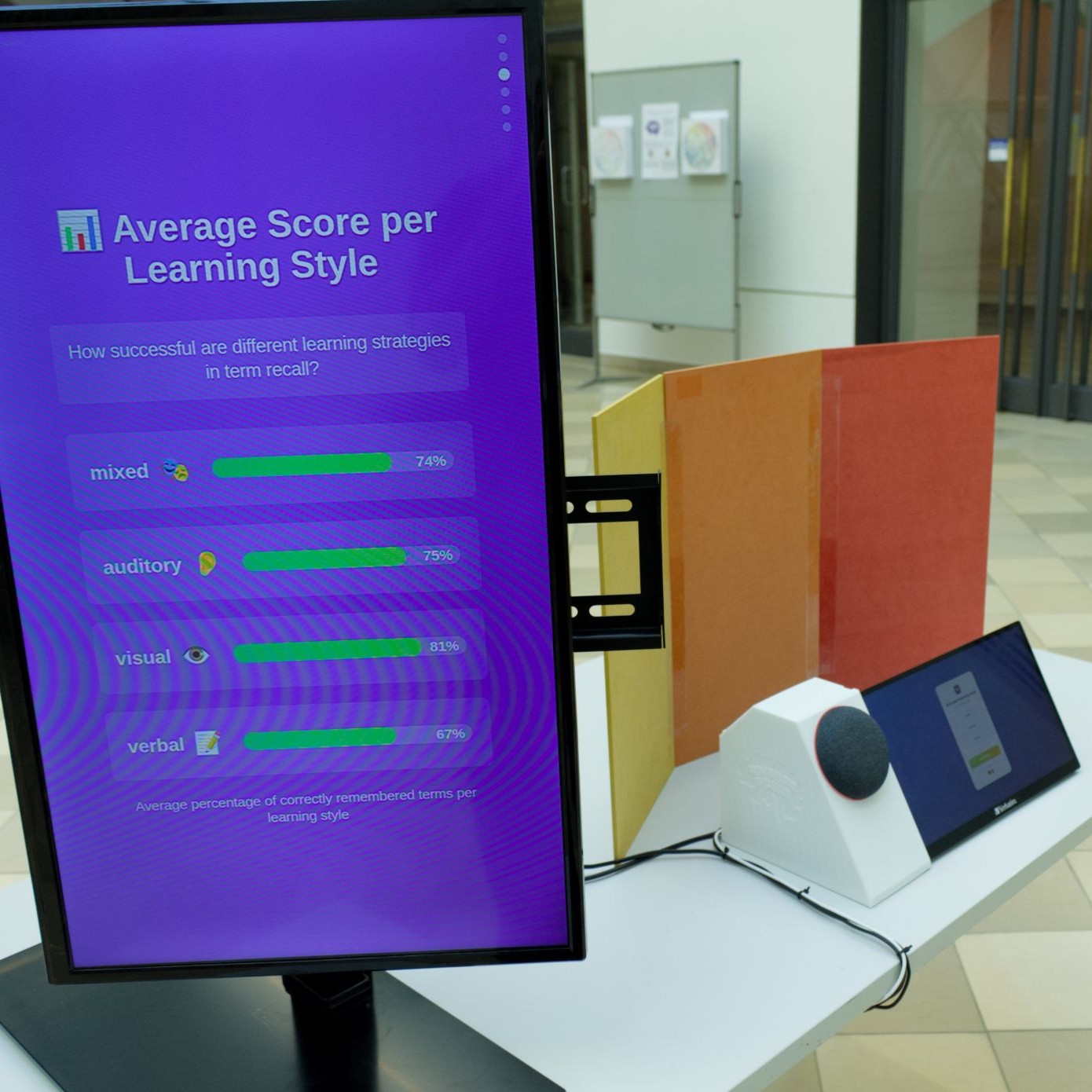
The idea that we can change simple things and achieve great impact has always been popular among people. One great example of such thinking are the different learning modalities.
These can be understood as different learning methods such as seeing, reading and hearing. Popular opinion suggests that each person has their own style of learning which boosts performance when used correctly. Contrary to this belief research suggest that learning styles are primarily preferences of the learner which have little effect on the ability to memorize compared to the non-preferred styles of learning [1, 2].
Additionally there is benefit in learning in many learning styles and modalities at once. This quiz compares peoples perfomances when using their preferred style of learning as well as all modalities (seeing, reading and hearing) at once.
Can we replicate these study results with a simple quiz? Participate here and find out!
References:
[1]: Testing the ATI hypothesis: Should multimedia instruction accommodate verbalizer-visualizer cognitive style? Author links open overlay panel by Laura J. Massa, Richard E. Mayer 2005
[2]:: Willingham, D. T., Hughes, E. M., & Dobolyi, D. G. (2015). The Scientific Status of Learning Styles Theories. Teaching of Psychology, 42(3), 266-271. https://doi.org/10.1177/0098628315589505 (Original work published 2015)
Representing fMRI Activity
Sara Wesolek Neuroscience (FU Berlin)
Michael Migacev Computer Science (FU Berlin)
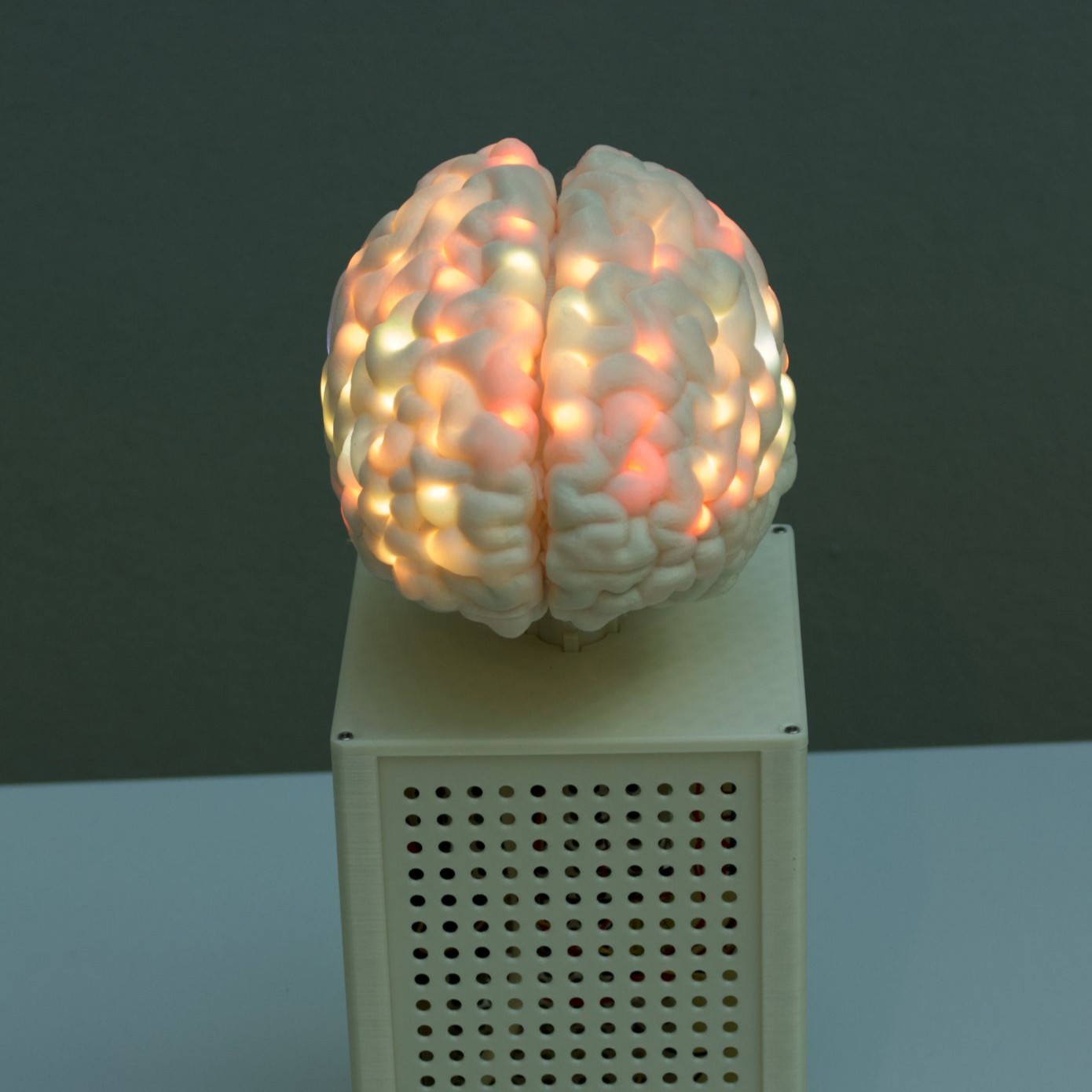
Traditionally, fMRI study results are presented as a figure or table. Especially as technology advances, the complexity of the data which must be illustrated increases.
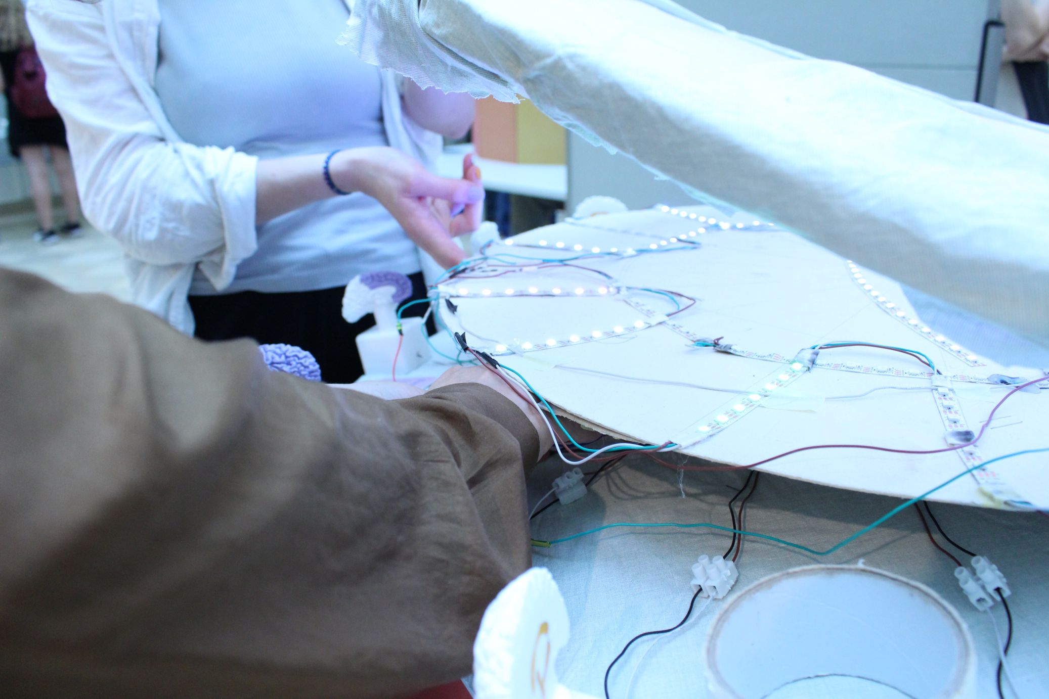
For example, Sara and their colleagues are working on a study which explores novel methods to record and analyze fMRI data resulting in ultra-high temporal resolution. This transforms the commonly seen 3D data into 4D data — activity patterns in three spatial dimensions across time. But how can these complex results be visualized?
While investigating the limitations of 2D paper and experimenting with modern 3D printing we have come up with an intuitive way to view such study results. 3D printing a hollow brain allowed us to attach LEDs along the shell. After cartographing each of the individual 204 LEDs, the lights can be individually addressed to display real study results — even complex data over time — on a 3D brain.
We hope to make neuroscientific research just a little more tangible with this project.
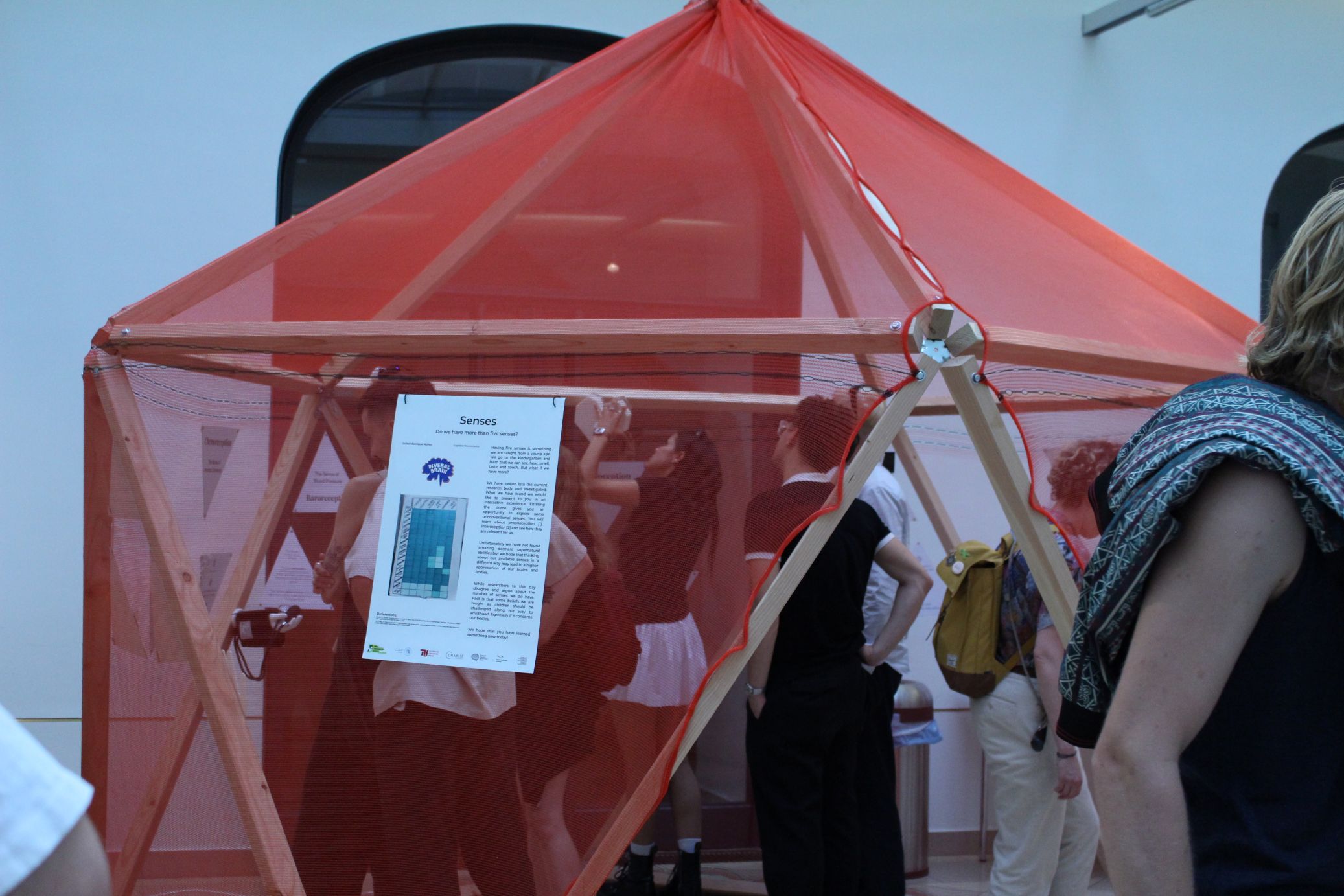
Senses
Luisa Manrique Núñez Cognitive Neuroscience (HU Berlin)
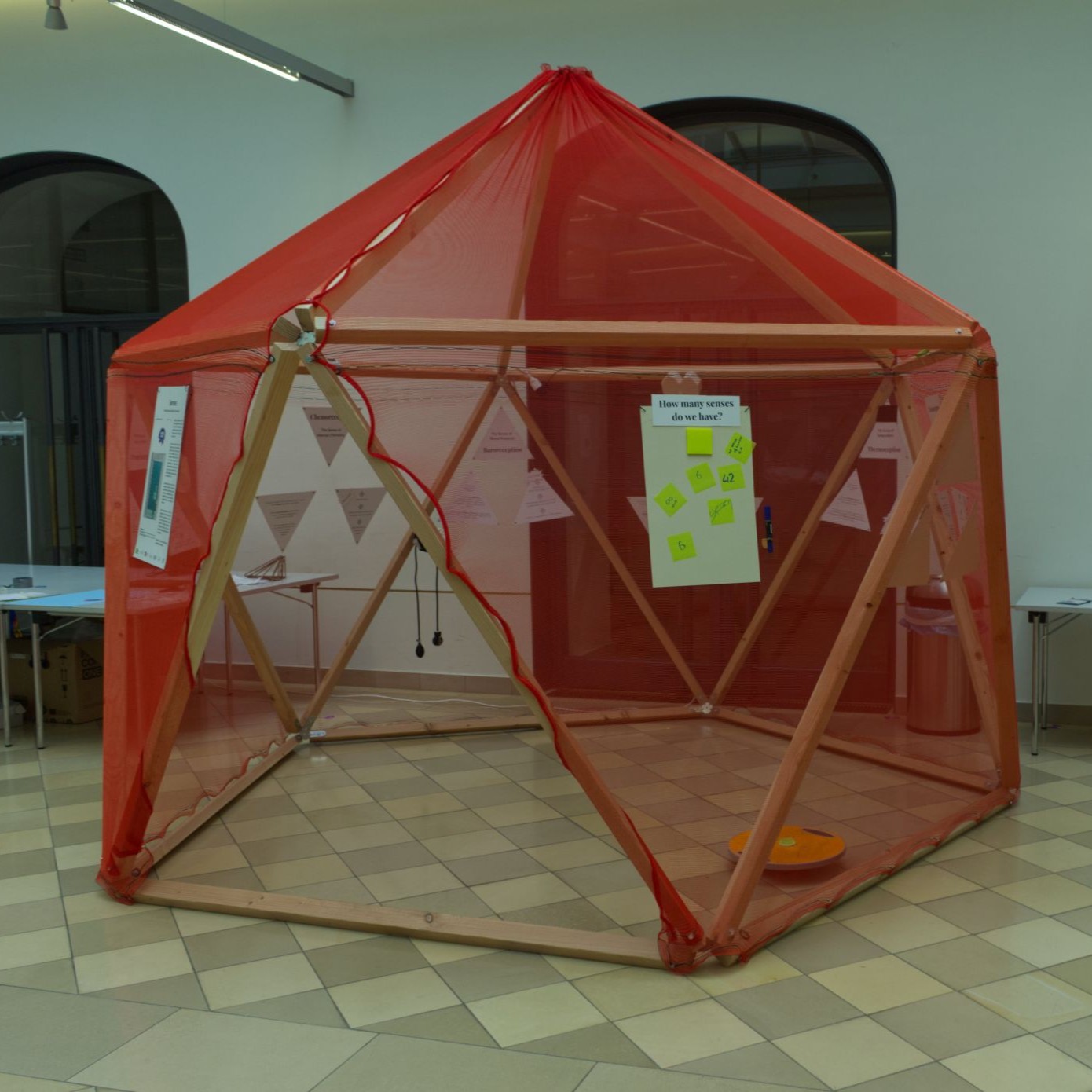
Having five senses is something we are taught from a young age. We go to the kindergarden and learn that we can see, hear, smell, taste and touch. But what if we have more?
We have looked into the current research body and investigated. What we have found we would like to present to you in an interactive experience. Entering the dome gives you an opportunity to explore some unconventional senses. You will learn about proprioception [1], interoception [2] and see how they are relevant for us.
Unfortunately we have not found amazing dormant supernatural abilities but we hope that thinking about our available senses in a different way may lead to a higher appreciation of our brains and bodies.
While researchers to this day disagree and argue about the number of senses we do have. Fact is that some beliefs we are taught as children should be challenged along our way to adulthood. Especially if it concerns our bodies.
We hope that you have learned something new today!
References:
[1]: Qin, Z. (2024). Proprioception. In: Kan, Z. (eds) The ECPH Encyclopedia of Psychology. Springer, Singapore. https:/ /doi.org/10.1007/978-981-97-7874-4_1246
[2]:: Craig, A. How do you feel? Interoception: the sense of the physiological condition of the body. Nat Rev Neurosci 3, 655—666 (2002). https://doi.org/10.1038/nrn894
Structural Connectivity
Carla Fillbrandt Psychology (FU Berlin)
Alena Beliaeva Veterinary Medicine (FU Berlin)
Zimo Gerlinger Psychology (FU Berlin)
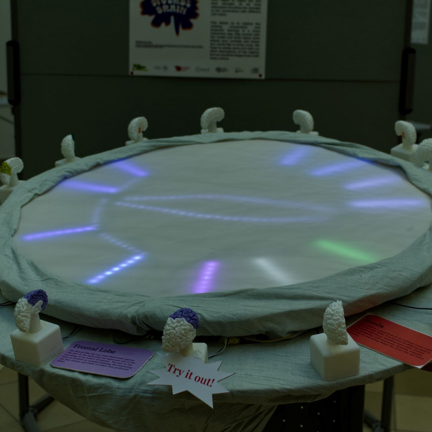
Current research is focusing on regions of the brain and analysing their function in relation to certain tasks. A lot of people overlook that these regions are connected not only to the regions next to them but also with a network of white matter lying underneath the grey matter.
We have followed a recent study which investigated structural connectivity[1] between the brain regions. Using that we have laid out an LED circle which represents connected regions. The buttons represent different regions in the left and right brain halves. The buttons are colored the show which regions they represent. Pressing the buttons reveals the connections between the regions. Not only do the connection vary from region to region but also their strength, so we have attempted encoding the strength of the connections with different LED colors.
This allows us to explore the existing connections and empower learning in a more interactive way. Play around with the model and think about what piques your curiosity and where you would like to know more. We hope to satisfy some of it with our short descriptions of the regions but we would be happy if you walk away curious.
References:
[1]: Škoch, A., Rehák Bučková, B., Mareš, J. et al. Human brain structural connectivity matrices—ready for modelling. Sci Data 9, 486 (2022). https://doi.org/10.1038/s41597-022-01596-9[1]: oinmckls am NJONCSAN cosd onk oincomlk nm aoi x20129 f sdjcn2981 10984u sdifj 193 3321423
Printing a Brain
Michael Migacev Computer Science (FU Berlin)
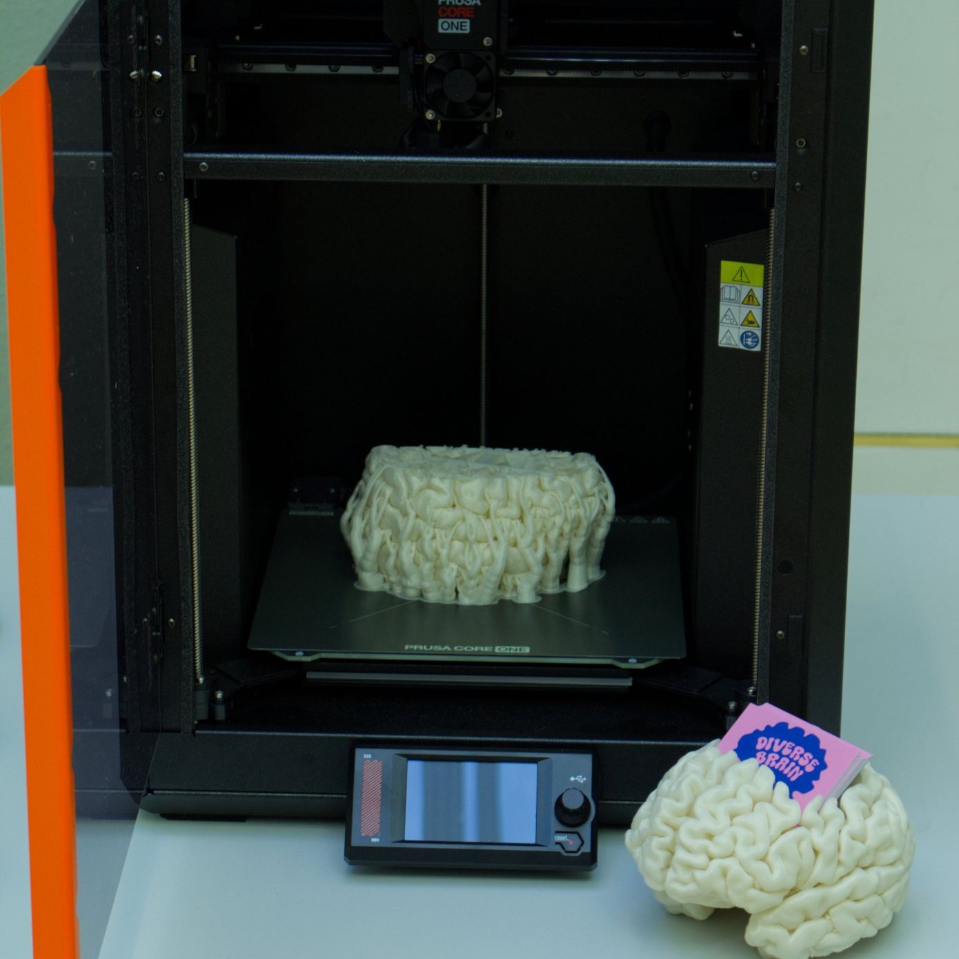
Modern MRI scanners are amazing. We can detect and distinguish matter within the millimeter. Combining this with common analysis methods let‘s us extract a 3D surface of the brain.
We encourage you to walk around and to see how different our brains look. The folds can almost be seen as a personal fingerprint. Although they all have a different shape researchers were unable to f ind corrrelation in function to the shape of the brain contrary to the once popular and luckily outdated belief of phrenology.
We have looked at all potential methods to generate the 3D surface and compared their results. We have finetuned the manual postprocessing which enables us to print the brains. Our code and video tutorial can be found at [1]
Want to get your own brain printed? We are currently working out a way to print your brain for you. If you would be interested make sure to write down your eMail on the note or shoot us an eMail at:
micha.migacev@googlemail.com
We will notify you once we are ready.
References:
[1]: https://github.com/printyourbrain/3DPrintYourBrain
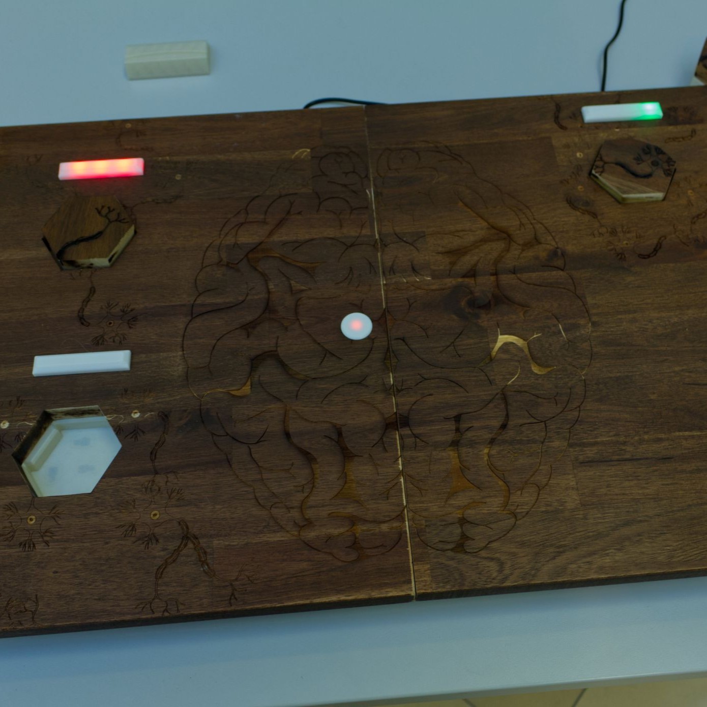
Left & Right Brain
Alexandra Groß Psychology (FU Berlin)
Andre Sebayang Psychology (FU Berlin)
Ahmed Abdelfatah Computational Neuroscience (TU Berlin)
Darvin Hassan Psychology (FU Berlin)
Michael Migacev Computer Science (FU Berlin)
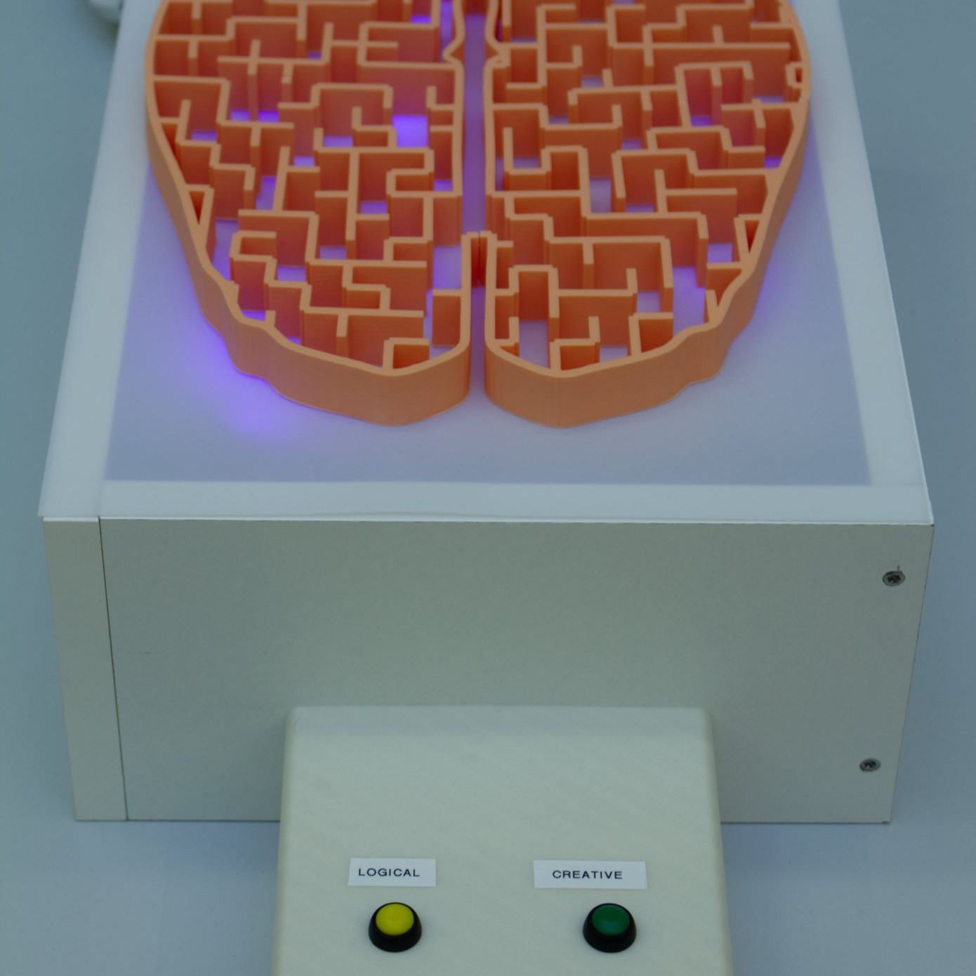
The brain can be described as a maze, in which certain paths are taken in certain situations. Unfortunately, there are many misconceptions about these " paths ", i.e. brain activity patterns, inspired by popular media, where thought-experiments often turn into misinformation.
The goal of our project was to debunk one such misconception: to show that being creative does not equal having a dominant "right brain", and being better at math does not equal having a dominant "left brain". Our interactive model, representing the activity patterns for creativity and math based tasks in an artistic interpretation. As you push the buttons of our model, you will see that the patterns are randomized and show little preference to specific brain halves. Although it is an artistic interpretation research suggests that complex mechanism such as logic or creativity are not bound to individual brain halves.
Instead, the activity patterns are most often divided on both brain hemispheres, though one hemisphere might be activated more strongly (Arsalidou et al., 2018; Bashwiner et al., 2020; Shah et al., 2013; Wang et al., 2022). Also, patterns vary from person to person (Finn et al., 2015). Similar things can be said about other brain functions, such as visual, auditory and emotional processing (Alho et al., 2014; Fusar-Poli et al., 2009, Li et al., 2022).
References:
[1]: Alho, K., Rinne, T., Herron, T. J., & Woods, D. L. (2014). Stimulus-dependent activations and attention-related modulations in the auditory cortex: a meta-analysis of fMRI studies. Hearing research, 307, 29-41.
[2]:: Arsalidou, M., Pawliw-Levac, M., Sadeghi, M., & Pascual-Leone, J. (2018). Brain areas associated with numbers and calculations in children: meta-analyses of fMRI studies. Dev. Cogn. Neurosci. 30, 239—250.
[3]:: Bashwiner, D. M., Bacon, D. K., Wertz, C. J., Flores, R. A., Chohan, M. O., & Jung, R. E. (2020). Resting state functional connectivity underlying musical creativity. NeuroImage, 218, 116940.
[4]:: Finn, E. S., Shen, X., Scheinost, D., Rosenberg, M. D., Huang, J., Chun, M. M., ... & Constable, R. T. (2015). Functional connectome f ingerprinting: identifying individuals using patterns of brain connectivity. Nature neuroscience, 18(11), 1664-1671.
[5]:: Fusar-Poli, P., Placentino, A., Carletti, F., Landi, P., Allen, P., Surguladze, S., ... & Politi, P. (2009). Functional atlas of emotional faces processing: a voxel-based meta-analysis of 105 functional magnetic resonance imaging studies. Journal of psychiatry and neuroscience, 34(6), 418-432.
[6]:: Li, X., O‘Sullivan, M. J., & Mattingley, J. B. (2022). Delay activity during visual working memory: A meta-analysis of 30 fMRI experiments. Neuroimage, 255, 119204.
[7]:: Shah, C., Erhard, K., Ortheil, H. J., Kaza, E., Kessler, C., & Lotze, M. (2013). Neural correlates of creative writing: an fMRI study. Human brain mapping, 34(5), 1088-1101.
[8]:: Wang, L., Li, M., Yang, T., Wang, L., & Zhou, X. (2022). Mathematics meets science in the brain. Cerebral Cortex, 32(1), 123-136.
Sleeping and Memory
Filippo Ghirardini University of Pisa (FU Berlin)
Viola Zeoli University of Trento (FU Berlin)
Michael Migacev Computer Science (FU Berlin)
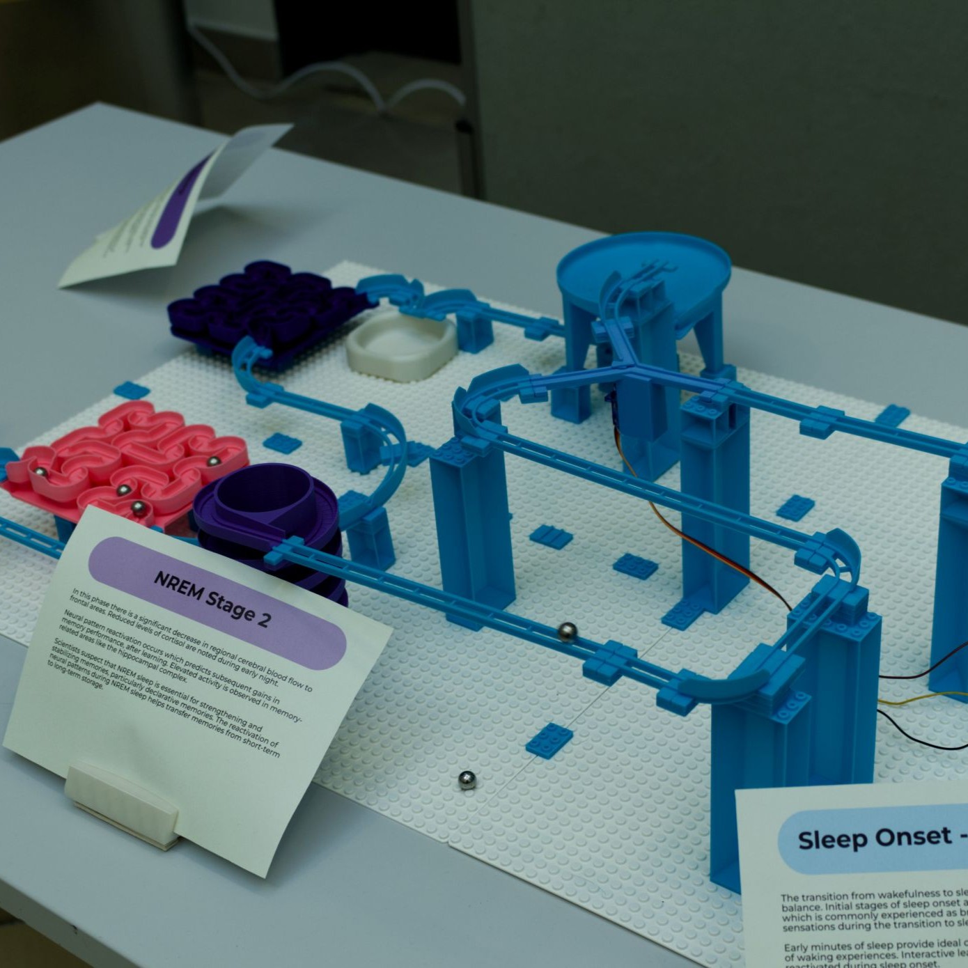
Sleeping is a natural human function. We are aware that when we sleep we rest and recover physically. Rarely can we appreciate or comprehend how sleep benefits our brain in many aspects. We will focus on the memory consolidation that happens during our sleep.
Researchers have determined that sleep is responsible for long term memory formations. They suggest these mechanisms to be at work:
1. hippocampal replay that, by replaying parts of our memories to us foster memorization
2. brain oscillations hallmarking slow-wave and rapid-eye movement sleep that provide mechanisms for regulating both information flow across distant brain networks and local synaptic plasticity;
3. qualitative transformations of memories during consolidation systems resulting abstracted, in gist-like representations. [2]
While these abstract concepts are difficult to grasp we have looked into how each of our sleep phases affects our memory formation. Try our accompanying marble run to learn more!
References:
[1]: Carskadon, Mary & Dement, William. (1989). Normal Human Sleep: An Overview. Principles and Practice of Sleep Medicine. M.H. Kryger (Ed.). W.B. Saunders, Philadelphia. 3-13.
[2]:: Klinzing, J.G., Niethard, N. & Born, J. Mechanisms of systems memory consolidation during sleep. Nat Neurosci 22, 1598—1610 (2019). https://doi.org/10.1038/s41593-019-0467-3
Global Integration Under Psilocybin
Kasey Devitt Cognitive Neuroscience (FU Berlin)
Psychedelics are being investigated for their therapeutic benefits. In neuroscience the study of psychedelics has revealed many insights into how these substances may alter brain connectivity. This model "Global integration Under Psilocybin," aims to visually represent the shift in brain region connectivity under the influence of psilocybin.
For many years it was thought that psychedelics primarily deactivated the medial prefrontal cortex (mPFC), a major node of the Default Mode Network (DMN). The DMN is a network of brain regions that are generally active when the brain is at rest. This led to the misconception that psychedelics simply "turn off" certain parts of the brain.
Recent research has corrected this. Rather than deactivating the mPFC. Psychedelics such as psilocybin cause a shift from local to global connectivity. This means that brain regions, including the mPFC, become more interconnected with other parts of the brain, leading to a state of increased global integration.
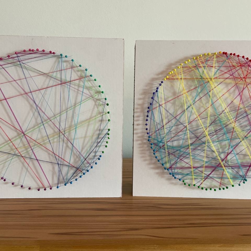
In our model, each color represents a particular area of the brain, and each pattern of connectivity represents a functional brain network, such as the Default Mode Network (DMN) or the Salience Network. The two circular diagrams in the artwork illustrate the difference in connectivity patterns.
Placebo
In the placebo condition, local connectivity is strong, and global connectivity is sparse. The strings between pins of the same color are more numerous, emphasizing the localized connections within specific brain regions. This represents the typical state of the brain when not under the influence of psychedelics, where functional networks like the DMN operate in a more isolated manner.
Psilocybin
Under the influence of psilocybin, local connectivity is disrupted, as illustrated by the lack of strings between pins of the same color. Instead, global connectivity is enhanced, with a significant increase in strings connecting nodes of different colors. This visual representation highlights the shift to global integration, where brain regions that were previously less connected now communicate more freely, leading to a more interconnected brain state.